Circle TIRF/SAIM galvo control
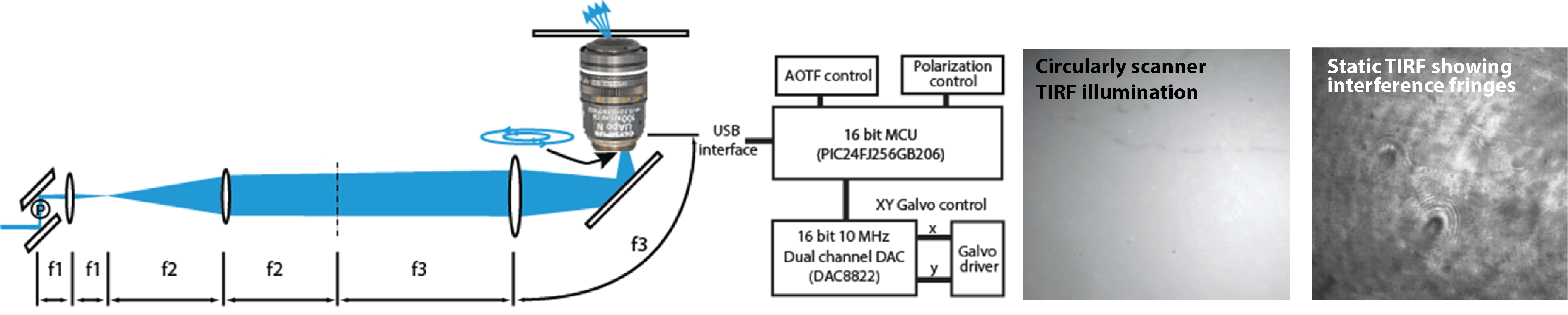
Our current scancard was originally designed for our LSM systems, but also can be used for circularly
scanned TIRF (also called azimuthally scanned TIRF) in which the focused excitation beam is scanned in a circle in the periphery
of the back focal plane of the objective lens. This achieves a more uniform TIRF illumination profile by averaging over the
interference patterns often seen in the conventional TIRF illumination field. The “circle scan” PIC firmware scans 32 X,Y points
to create the circle.
In a collaboration with the Paszek Lab we also applied the hardware to improve scanning angle interference microscopy (SAIM).
Using a series of sequential circular scan diameters and EMCCD synchronization we sped up and automated SAIM imaging.
A Biophysics PhD student in the Paszek lab (Marshal Coleville) worked with our lab to modify the scancard further
to create a smoother circular scan by adding a function generator IC to the scancard PCB. The complete schematics
and firmware files are available for this design on GitHub.
Colville M, Park, S, Zipfel, WR, Paszek, MJ “High-speed device synchronization in optical microscopy
with an open-source hardware control platform” Sci Rep 9, 12188 (2019).
Colville M, Park S, Singh A, Paszek M, Zipfel WR. Azimuthal Beam Scanning Microscope Design and Implementation for Axial Localization with Scanning Angle Interference Microscopy. Methods Mol Biol. 2022; 2393:127-152. doi:10.1007/978-1-0716-1803-5_7. PMID: 34837177.
Colville M, Park S, Singh A, Paszek M, Zipfel WR. Azimuthal Beam Scanning Microscope Design and Implementation for Axial Localization with Scanning Angle Interference Microscopy. Methods Mol Biol. 2022; 2393:127-152. doi:10.1007/978-1-0716-1803-5_7. PMID: 34837177.